
Heart – Anatomical Overview
The heart as the centre of the circulatory system is located anterior to the vertebral column and posterior to the sternum.
It is enclosed in a double-walled sac called the pericardium. The pericardium’s outer wall is called the parietal pericardium and the inner one the visceral pericardium. Between them there is some pericardial fluid which functions to permit the inner and outer walls to slide easily over one another with the heart movements. Outside the parietal pericardium is a fibrous layer called the fibrous pericardium which is attached to the mediastinal fascia. This sac protects the heart and anchors it to the surrounding structures.
The outer wall of the human heart is composed of three layers; the outer layer is called the epicardium, or visceral pericardium since it is also the inner wall of the pericardium. The middle layer is called the myocardium and is composed of contractile cardiac muscle. The inner layer is called the endocardium and is in contact with the blood that the heart pumps. Also, it merges with the inner lining (endothelium) of blood vessels and covers heart valves.

The human heart has four chambers, two superior atria and two inferior ventricles. The atria are the receiving chambers and the ventricles are the discharging chambers. During each cardiac cycle, the atria contract first, forcing blood that has entered them into their respective ventricles, then the ventricles contract, forcing blood out of the heart. The pathway of the blood consists of a pulmonary circuit and a systemic circuit which function simultaneously. Deoxygenated blood from the body flows via the vena cava into the right atrium, which pumps it through the tricuspid valve into the right ventricle, whose subsequent contraction forces it out through the pulmonary valve into the pulmonary arteries leading to the lungs. Meanwhile, oxygenated blood returns from the lungs through the pulmonary veins into the left atrium, which pumps it through the mitral valve into the left ventricle, whose subsequent strong contraction forces it out through the aortic valve_ to the aorta leading to the systemic circulation.

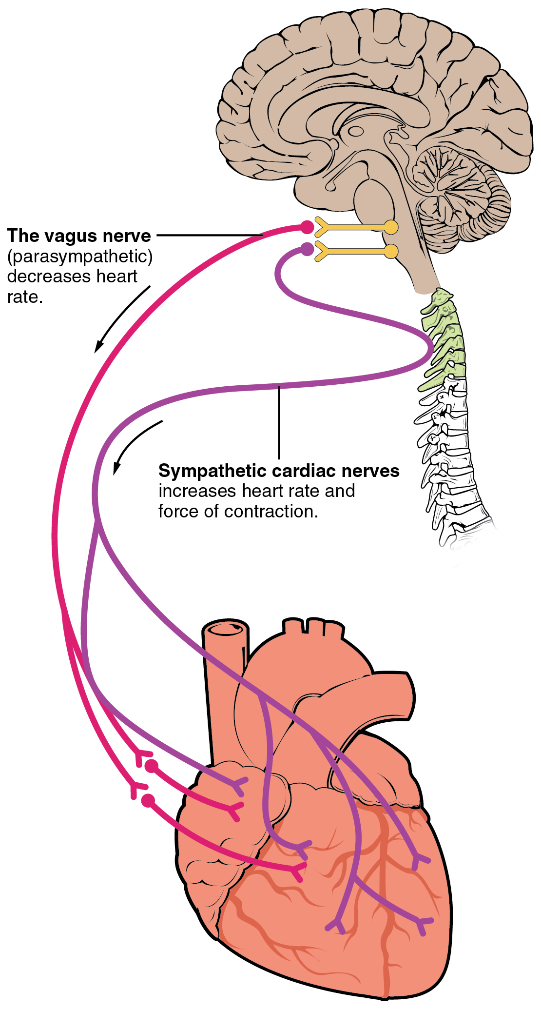
The myocardium contracts after stimulation. Under normal conditions, electrical activity is spontaneously generated by the SA node, the physiological pacemaker, and is closely tied to sympathetic discharge. This electrical impulse is propagated throughout the right atrium, and through Bachmann’s bundle to the left atrium, stimulating the myocardium of the atria to contract. As the electrical activity is spreading throughout the atria, it travels via specialized pathways, known as internodal tracts, from the SA node to the AV node. The AV node functions as a critical delay in the conduction system. Without this delay, the atria and ventricles would contract at the same time, and blood wouldn’t flow effectively from the atria to the ventricles. Second order electrical control of the myocardium is closely tied to parasympathetic influence from the spinal vertebral ganglia and vagus nerves.

graphic: Wikipedia, Patrick J Lynch
From an evolutionary point of view, the heart has made a turn so that curiously, in the Plexus Cardiacus (autonomous nerve system or XIII brain nerve), fibres are running together which relate both to the brain stem relay crossing over, and from cerebral cortex relays not crossing over. So, the relay for the sinus node that stimulates heart rhythm via the right atrium, lies in the left brain stem.
Brain

graphic: Phil Schatz

graphic: McGraw-Hill Companies, 2002
Relays
The Plexus Cardiacus contains fibres related to following relays:
Cerebral Medulla (-/+) medial of “Throacic region” left & right, for Endocardium & Valves and Ventricular Myocardium

Midbrain (+/-) fronto-lateral left & right, for Atrial Myocardium and stimulation via sinoatrial node (heart rhythm)
compare Brain Stem 
Cerebral Cortex (-/+) insular region medio-lateral left & right, for Coronary Arteries and Veins and AV Node (pulse regulation, bradycardia)

Cerebellum (+/-) medial for Pericardium & Epicardium
